
Obtaining accurate and precise results is crucial in bio-medical research, and to achieve this we often turn to advanced technology. A key player in improving microscopy, which is a fundamental tool in this field, is the Microscope-Cockpit software. This software makes custom-built microscopes much more powerful and easier to use. One of the main reasons for its effectiveness is the integration of a Red Pitaya device, a flexible and powerful single-board computer. This article investigates how Red Pitaya is helping to change the game in microscopy, making tasks that were once complex and time-consuming simpler and more efficient.

Microscope-Cockpit for Beginners
Before diving into the technicalities, it's essential to understand what Microscope-Cockpit is and why it's a game-changer for researchers. At its core, Microscope-Cockpit is a software platform designed to operate complex microscopy systems—imagine having a sophisticated camera system that can not only take detailed pictures of microscopic entities but also control the light, focus, and movement automatically to capture the best images possible. Developed with the Python programming language, it's both flexible and powerful, allowing researchers to customize their experiments and integrate new tools and algorithms as needed. Whether you're studying the structure of a virus or monitoring live cells, Microscope-Cockpit streamlines the process, making advanced imaging techniques more accessible to scientists of all skill levels.

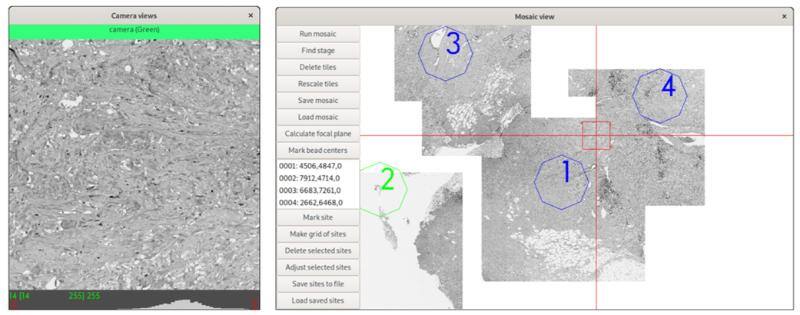
Figure 1: The main Cockpit Graphical User Interface (GUI) components: (a) The main window provides quick access to several functions and shows the status of all the devices, as well as the channel buttons to load predefined configurations. (b) The macro stage window provides an overview of the position of all stages, including nested stages and the Z position. (c) The camera view shows the last image taken from each active camera and the related histograms. (d) The mosaic view displays all mosaic images taken and the saved locations, and dramatically eases navigation.
Red Pitaya in Microscope-Cockpit
Microscope-Cockpit, developed by Mick A. Phillips and his team, is a Python-based, open-source graphical user interface (GUI) designed for the precise control of both simple and elaborate custom-built microscope systems. Among its many innovative features, the integration of a Red Pitaya device is particularly noteworthy. Serving as a high-performance hardware timing device, Red Pitaya ensures that the Microscope-Cockpit can conduct time-critical operations with unparalleled precision.
The use of Red Pitaya within Microscope-Cockpit is a testament to its reliability and efficiency in facilitating complex experiments. Whether coordinating the actions of multiple hardware devices or achieving synchronized analog voltages for structured illumination, the Red Pitaya enables the software to maintain consistent and accurate control throughout the imaging process.
Key Applications of Red Pitaya in Zaber Imaging and DeepSIM
The integration of Red Pitaya into the Zaber Imaging and DeepSIM systems highlights its critical role in advancing microscopy technology. This versatile single-board computer significantly enhances the functionality of these systems by managing the precise timing and synchronization needed for complex imaging tasks.
Zaber Imaging System
In the Zaber Imaging System, Red Pitaya optimizes the control of LED light sources for fluorescence imaging, enabling rapid switching between different wavelengths without manual filter changes. This capability is crucial for effective multi-channel imaging, allowing researchers to capture dynamic biological processes with high precision and minimal delay. The integration of Red Pitaya ensures that the compact, high-resolution Ximea camera operates efficiently within the space constraints of modern laboratories, making advanced imaging more accessible.


Figure 2: The Zaber microscope: a) An image of the system, showing its compact size with a 300×450 mm footprint. (b) A three colour image showing the nucleus stained with DAPI in blue, the actin in green and mitochondria stained with MitoTracker Red CMXRos in Red. Scale bar 5 µm.
DeepSIM System
For the DeepSIM system, the Red Pitaya board synchronizes laser illumination with the spatial light modulator (SLM) and deformable mirror (DM) to execute structured illumination microscopy (SIM) combined with adaptive optics (AO). This precise control is essential for achieving super-resolution and correcting optical aberrations in real time, which is particularly important for deep tissue imaging. The Red Pitaya’s role is vital in live imaging experiments, where capturing fleeting cellular processes with clarity can provide deep insights into biological mechanisms.

Figure 3: Example data from the DeepSIM system using Cockpit to image deep (<20µm) in Drosophila neuro-muscular junctions: (a) System view from the stage. (b) Main Cockpit window with the additional controls for the SLM and AO devices. (c) Image of individual synapses at the Drosophila neuro-muscular junction using AO SIM imaging, with Cy3 labelled HRP antibody labelling the neuronal membrane in red and Brp: GFP in the synapse in green. (d) Zoomed in region of (c), the scale bars are 2µm.
In both Zaber Imaging and DeepSIM, the use of a Red Pitaya board facilitates a higher level of detail and efficiency in microscopy studies. It enables sophisticated imaging techniques that combine super-resolution with real-time optical corrections, pushing the boundaries of bio-medical research. By simplifying the control of complex imaging processes, the Red Pitaya board not only enhances operational efficiency but also improves the scientific outcomes of these advanced microscopy systems.
Conclusion
The integration of a Red Pitaya board within Microscope-Cockpit has profound implications for bio-medical science. It not only makes access to advanced microscopy techniques more widespread, but also significantly enhances the quality and efficiency of scientific research. By enabling precise control and synchronization of complex imaging processes, researchers can explore cellular structures and dynamics with an unprecedented level of detail, paving the way for groundbreaking discoveries in bio-medical science.
In summary, the Microscope-Cockpit, empowered by the integration of Red Pitaya, represents a significant advance in microscopy control software. Its ability to handle complex, time-sensitive imaging tasks with ease and precision makes it a pivotal tool in the arsenal of bio-medical researchers. As we continue to unravel the mysteries of the microscopic world, the collaboration between Red Pitaya and Microscope-Cockpit will undoubtedly play a crucial role in shaping the future of bio-medical science.


