Bone Fracture Detection Using Electrical Impedance Tomography with a Red Pitaya STEMlab
-
Posted by
 Red Pitaya Technical Editorial Team
, November 7, 2024

Bone fractures are a common medical issue, often resulting from accidents, osteoporosis, or bone cancer. Traditional methods for detecting fractures, such as X-rays, MRI, and CT scans, while effective, come with drawbacks like high costs and potential radiation exposure. Enter Electrical Impedance Tomography (EIT) – a promising, non-invasive imaging technique that leverages electrical impedance distribution to visualize internal body structures. In this blog post, we'll explore how the Red Pitaya STEMlab module plays a pivotal role in advancing EIT for bone fracture detection.
Understanding Electrical Impedance Tomography (EIT)
EIT is an imaging modality that maps the electrical impedance within body tissues. Different tissues exhibit varying impedance levels, allowing EIT to identify abnormalities like fractures based on these variations. For instance, fractured bone tissue often shows increased blood flow and local edema, leading to higher conductivity in the affected area. By detecting these changes in conductivity, EIT can effectively locate and characterize bone fractures.
Why Use a Red Pitaya STEMlab?
The Red Pitaya STEMlab is a versatile, open-source measurement and control platform known for its affordability and flexibility. It integrates multiple functions such as a voltage generator, oscilloscope, and multiple input/output channels, making it an ideal controller for EIT systems. Its digital and analog capabilities allow seamless signal generation, data acquisition, and real-time processing, essential for accurate impedance measurements and image reconstruction.
System Design and Implementation
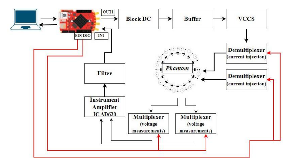
The EIT system developed for bone fracture detection comprises both hardware and software components:


Hardware Components:
- Red Pitaya STEMlab Module: Acts as the central controller, generating AC voltage signals and capturing voltage measurements.
- Voltage Controlled Current Source (VCCS): Ensures a constant and stable current injection into the bone phantom.
- Multiplexer/Demultiplexer Circuits: Manage the injection of current and measurement of voltage across multiple electrodes.
- Amplifiers and Filters: Enhance signal quality by amplifying weak signals and filtering out noise.
Software Components:
MATLAB plays a crucial role in controlling the Red Pitaya module. It is utilized for generating voltage signals, managing data acquisition, and processing the collected data. The integration of MATLAB with the Red Pitaya allows for precise control over the signal generation and data processing workflows. Furthermore, the Electrical Impedance and Diffuse Optical Reconstruction Software (EIDORS), a dedicated MATLAB toolbox, is employed for image reconstruction from the impedance measurements. EIDORS facilitates the transformation of raw impedance data into meaningful tomographic images, enabling the clear visualization of bone fractures.
Creating the Bone Phantom
To simulate bone and soft tissue, a 3D-printed polylactic acid (PLA) bone phantom was developed. This phantom mimics the electrical properties of real bone and was embedded in a water-filled acrylic chamber representing soft tissue. Sixteen electrodes were strategically placed around the phantom to facilitate current injection and voltage measurement, enabling comprehensive impedance mapping.
Data Acquisition and Analysis
The data acquisition process involved injecting a controlled current into the bone phantom through adjacent electrodes and measuring the resulting voltage. This process was repeated across all electrode pairs, generating a comprehensive set of impedance measurements. The collected data were then processed using EIDORS to reconstruct tomographic images, highlighting differences in electrical impedance indicative of fractures.


Results
The EIT system successfully distinguished between normal and fractured bone phantoms. Reconstructed images showed clear conductivity variations, with fractured areas exhibiting higher conductivity due to increased blood flow and edema. The use of Red Pitaya ensured stable signal generation and accurate data acquisition, underscoring its effectiveness in medical imaging applications.
Conclusion
Integrating a Red Pitaya STEMlab into an EIT system is a cost-effective and reliable solution for bone fracture detection. This approach not only reduces the risks associated with traditional imaging techniques but also offers portability and ease of use, making it a valuable tool in medical diagnostics. Future enhancements could include using more bone-like materials for phantoms or exploring advanced image reconstruction algorithms to further improve detection accuracy.
About the Red Pitaya Team
The Red Pitaya Technical Editorial Team is a cross-functional group of technical communicators and product specialists. By synthesizing insights from our hardware developers and global research partners, we provide verified, high-value content that bridges the gap between open-source innovation and industrial-grade precision.
Our mission is to make advanced instrumentation accessible to engineers, researchers, and educators worldwide.


